Media Summary: MIT researchers led by Ed Boyden have invented a new way to visualize the nanoscale structure of the brain and other tissues. Heather introduces us to the concepts and practicalities of performing ... have longer imaging times and require more expertise
Expansion Microscopy Explained - Detailed Analysis & Overview
MIT researchers led by Ed Boyden have invented a new way to visualize the nanoscale structure of the brain and other tissues. Heather introduces us to the concepts and practicalities of performing ... have longer imaging times and require more expertise His group at MIT develops tools that enable the mapping of the molecules and wiring of the brain, the recording and control of its ... An alternative approach to the visualization of subcellular microstructures, which relies on “enlarging” the sample, rather than ... Hello my name is maddie and today we're going to talk about
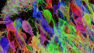
Did you know that you can literally expand biological samples to reveal hidden details? This is exactly what's possible with ... Edward Boyden is a Hertz Foundation Fellow and recipient of the prestigious Hertz Foundation Grant for graduate study in the ... Instructional video for Cell ExM (Expansion microscopy) Kit Brainbow-expressing mouse hippocampal brain circuitry, processed via the proExM form of Fei Chen co-invented a technique to beat the "diffraction limit" - the overlapping of waves of light (roughly a few hundred ... Flythrough of image data collected from mouse hippocampus, with neurons expressing Yellow Fluorescent Protein, showing both ...
Join Prof. Dr. Lorenz Sellin of the University Hospital of Düsseldorf as he presents on the topic: Imaging of renal proteins with ...